The medical physics team performs extensive simulation studies in both diagnostic and therapeutic fields. Two imaging modalities are mainly explored: Positron Emission Tomography (PET) for cancer diagnosis and Magnetic Resonance Imaging (MRI) for detection of heart anomalies. As for radiation therapy, the full chain of the clinical radiotherapy facility is simulated using Monte Carlo codes.
Nuclear Medicine:
Positron Emission Tomography is a nuclear imaging modality that uses small amounts of radioactive materials coupled to a biologically active molecule called radiotracers to explore human physiology. It is heavily used in clinical oncology for cancer diagnosis.
The radiotracer injected in the human body and absorbed by tissues of interest (mainly tumors) emits positrons that annihilate with electrons to produce a pair of photons, each of energy 511 keV, emitted in opposite directions. PET scanners are designed to detect coincident photons and localize their point of emission along the coincidence line called the line of response (LOR). The source of the photons which corresponds to the annihilation site is needed to create a 3D distribution of the radiotracer in the target organ.
We use Monte Carlo based software to model and validate clinical PET scanners by comparing our simulation results to clinical measurements. We extend our simulation studies in this field to include modeling and optimization of Total Body PET scanners, a very promising project that we collaborate on with our international partners.
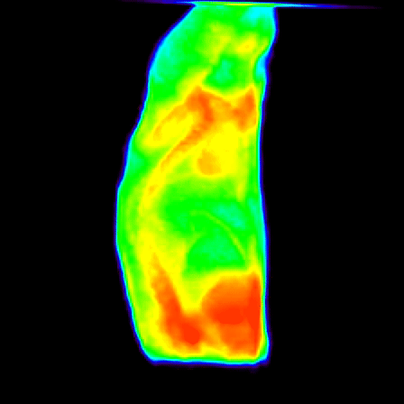
MRI imaging:
We are using MRI image analysis for Visual Analysis of Regional Myocardial Motion. This helps detect regional anomalies in the myocardial motion of the left ventricle (LV) which are important biomarkers for several cardiac diseases.
The group is also working on algorithms for fast MRI analysis based on parallel programming and new accelerator technologies.
Radiation Therapy:
Radiation Therapy (RT) for cancer treatment is also explored to fully simulate the clinical linear accelerator that delivers a high energy beam of radiation to damage cancer cells, and to calculate the dose distribution needed for the Treatment Planning System (TPS).
ACK: all computational resources, 3D visualization tools and scientific software are provided by the Research Computing at TAMUQ.